This chapter introduces the Point Source Microscope (PSM) as a multipurpose optical metrology
instrument. For those already familiar with the PSM, many of the following concepts may seem
straightforward. However, for new users—or those with limited background in optics—the
operation and capabilities of the instrument are not immediately obvious. Even experienced users
may not fully appreciate certain aspects of its design.
The goal of this chapter is to present the optical functions of the PSM in a systematic way,
building an understanding of how its individual components work together to form a versatile
and precise alignment tool.
Figure 1 illustrates the principal optical paths within the PSM and provides a global view of the
instrument.

Fig. 1 Optical paths within the PSM
From this starting point, we examine each functional mode of the instrument in turn, to explain
the operation of each part of the instrument in terms of other familiar optical instruments.
PSM as a Telescope
When the infinite conjugate microscope objective is removed, the PSM functions as a telescope,
as shown in Fig. 2. In this configuration, light from a distant object is brought to focus on the
CCD camera. If the object were a star, the image formed would be a diffraction-limited spot.
Although the PSM is not typically used as a telescope, this configuration is essential for
instrument setup during its assembly. The tube lens (referred to here as the telescope objective) is
adjusted by observing a distant object and optimizing focus on the CCD. Once set, this
establishes the PSM as an infinite conjugate system.

Fig. 2 The PSM as a telescope focused at infinity when the microscope objective is removed
PSM as a Transmission Microscope
With the tube lens properly adjusted, an infinite conjugate microscope objective can be added,
converting the PSM into a transmission microscope (Fig. 3). When a transparent or translucent
sample is placed at the objective focus and illuminated externally, a magnified image is formed
on the CCD.
Fig. 3 The PSM as a transmission microscope
Due to the 100 mm effective focal length (EFL) of the PSM tube lens—approximately half that
of typical biological microscopes—the effective magnification is reduced accordingly. For
example, a 10× objective produces a field of view of approximately 1.2 mm on the sample.
While this mode is not a primary application of the PSM, it plays an important role in instrument
assembly and calibration.
PSM as a Collimator
Extending the telescope concept, the PSM can operate as a collimator by introducing an internal
point source (Fig. 4). In this configuration, light from a single-mode fiber is placed at the focal
plane of the objective, producing a collimated beam.
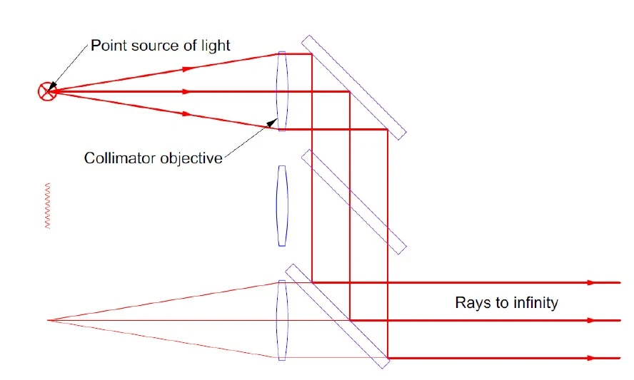
Fig. 4 The PSM as a collimator using an internal point source of illumination
A collimator is effectively a telescope operated in reverse: instead of forming an image of a
distant object, it projects a point source to infinity. This produces an artificial star, commonly
used in optical testing.
In the PSM, the collimated beam has a diameter of approximately 10 mm and exhibits a
Gaussian intensity distribution due to the single-mode fiber source. While this limits its
usefulness for conventional lens testing, it is sufficient for small-aperture applications.
Projection of a Spherical Wavefront
When a microscope objective is attached in the collimator configuration, the PSM projects a
spherical wavefront (Fig. 5), analogous to the output of an interferometer transmission sphere.
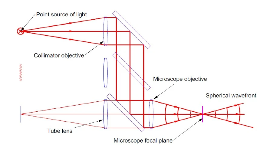
Fig. 5 The PSM as a spherical wavefront projector
At the focal plane, a diffraction-limited spot is formed, conjugate with the fiber source. The
numerical aperture (NA) and divergence of the beam are determined by the objective, allowing
control over the cone angle of illumination.
Establishing the Coordinate Origin of the PSM
A key capability of the PSM is the determination of a stable coordinate origin on the CCD.
When a matte surface is placed at the focal plane, the illuminated spot is imaged onto the CCD
via scattered light (Fig. 6). Importantly, transverse motion or tilt of the surface does not change
the position of the spot on the detector. This invariance establishes a fixed reference point.
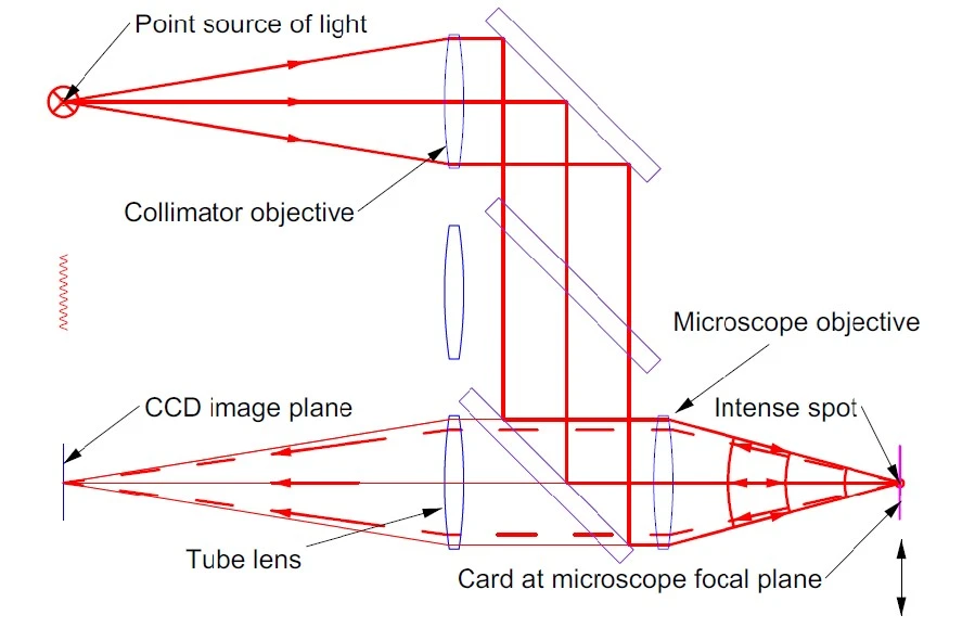
Fig. 6 The PSM viewing scattered light from a matte surface at the microscope objective focus
In practice, a smooth, specular surface is used, producing a well-defined spot that is easier to
centroid. Although using a specular surface is less intuitive to explain in terms of how the spot is
imaged, the result is the same as using a matt surface to get a Cat’s eye return.
Cat’s Eye Reflection
With a smooth surface, the reflection is specular and produces a Cat’s eye effect. This
phenomenon is illustrated schematically in Fig. 7.

Fig. 7 Cat’s eye reflection from a specular surface at normal incidence (left) and tilted (right)
When the surface is normal to the optical axis (lefthand view), the incident rays (red) are
reflected (green) where the angle of incidence equals the angle of reflection about the normal to
the surface and return to the detector. When the surface is tilted, the reflected light ray cone
rotates about the point of incidence, and some of the reflected light is vignetted, but the point of
incidence remains fixed. Consequently, the image on the CCD does not shift.
This property is used to define the reference position on the CCD. Once the Cat’s eye reflection
is centered, the system establishes a precise optical origin. All subsequent measurements are
made relative to this reference.
This process is analogous to boresighting a riflescope: once aligned, the system ensures that
outgoing light is centered on the reference axis to within micrometer precision.
Autostigmatic Operation
The Cat’s eye configuration introduces the concept of “auto” operation. In this regime, light
emitted by the instrument is reflected back into the objective along the same path as it was
emitted and is detected internally, forming the basis of an autostigmatic microscope.
Thus, with the microscope objective in place, the PSM operates as an autostigmatic microscope.
The term “stigma,” or point, from the Latin, refers to the point where a cone of rays converge.
Thus, the instrument combines autocollimation with finite-distance, or point, focusing to be an
autostigmatic microscope.
Reflecting Microscope Mode
The PSM also functions as a reflecting inspection microscope. An LED-illuminated diffuser
provides full-field illumination, allowing visualization of the sample (Fig. 8a). The internal point
source produces a localized Cat’s eye spot (Fig. 8b), and both sources can be used
simultaneously (Fig. 8c).
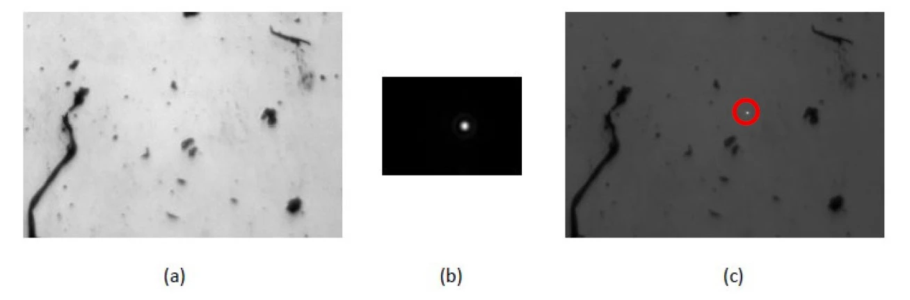
Fig. 7a. Sample illuminated with the LED source only where the FOV is 1 mm horizontally, Fig. 7b. Cat’s eye image off the sample using the point source where the FOV is 0.1 mm horizontally, and Fig. 7c. with the sample illuminated using both sources where the Cat’s eye spot is in the red circle. The electronic crosshair is not saved in the software, but the Cat’s eye spot in (c) is precisely centered in the crosshair as seen on the PSM monitor screen.
By aligning the imaging and illumination paths to be parfocal, the PSM ensures that both the
projected focused spot and the detected image share a common reference plane. This critical
property is adjusted during the instrument assembly. This dual illumination feature allows a
micron-scale spot to be positioned precisely on any feature within the field of view.
Use of External Sources
The internal light source can be replaced with an external fiber-coupled source via an FC/APC
connector (Fig. 9a). This enables operation over a wide wavelength range. A filter slot allows
wavelength-selective detection, enabling pump–probe or fluorescence-type measurements when
appropriate filters are used (Fig. 9b).

Conclusion
The Point Source Microscope integrates multiple optical functions—telescope, microscope,
collimator, and autocollimator—into a single instrument. Across these modes, it provides angular
sensitivity at the sub-arcsecond level and positional sensitivity at the sub-micrometer level.
The ability to use external light sources further expands its versatility, enabling applications
across a broad spectral range. While the standard configuration supports visible and near-infrared
operation, extended wavelength options allow the PSM to address specialized measurement
tasks. Together, these capabilities make the PSM a uniquely flexible, small footprint and
lightweight tool for optical alignment and metrology.